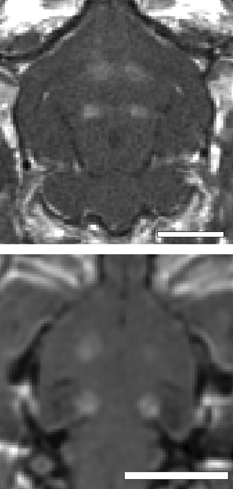
Figure 1.
Coronal contrast-enhanced T1-weighted images in the rabbit (top) and rat (bottom) brains showing the spatial location of ultrasound exposures and BBB disruption. The spacing between the four exposures was adjusted based on the size of the brain. All exposures were performed through intact skull.

