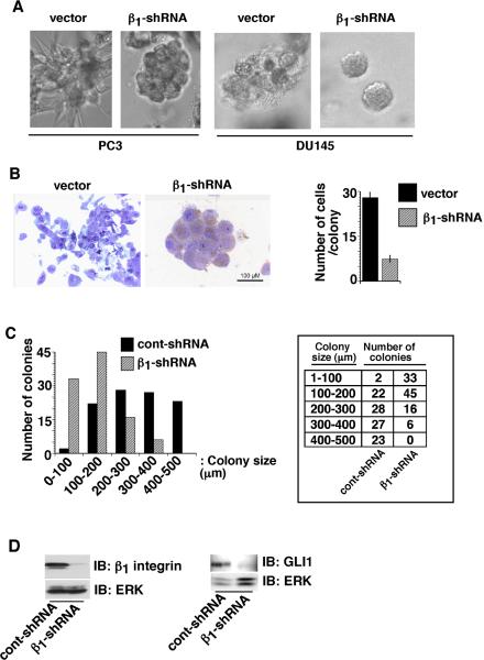
Fig. 2. β1 integrins reduce cell proliferation in 3-D cultures.
A: PC3-vector, PC3-β1-shRNA, DU145-vector and DU145-β1-shRNA transfectants were cultured in Matrigel. Colonies in 3-D cultures were observed under a phase contrast microscope and images were captured. B: PC3-vector and PC3-β1-shRNA transfectants cultured in Matrigel were fixed and sections were stained with 1% toluidine blue (left panels). Number of cells in each colony was counted and shown as average with SEM (n=20, right panel, p<0.00001). C: PC3-cont-shRNA and PC3-β1-shRNA transfectants were cultured in Matrigel for 12 days. Colonies in 3-D cultures were observed and the size of each colony was measured using a phase contrast microscope. Chi-square for linear trend with 1 degree of freedom is 73.462 (p<0.00001). D: PC3-cont-shRNA and PC3-β1-shRNA transfectants were cultured in Matrigel for 12 days. Cells were isolated from Matrigel using PBS-EDTA and then lysed. Lysates were separated and immunoblotted using Abs to β1 (C-18), GLI1 or ERK, as a loading control.

