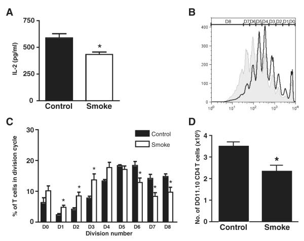
FIGURE 4.
Cigarette smoke exposure inhibits lung-derived DC induced T cell proliferation in vitro and in vivo. A, Newly migrated CD11c+/OVA+ lung-derived DCs were isolated from the TLNs of smoke-exposed and control mice 24 h following intratracheal injection of FITC-labeled OVA and cocultured with DO11.10 CD4+ T cells. IL-2 levels were measured in culture supernatants by ELISA. Data represent means ± SEM of triplicate culture wells, each assayed in duplicate. Statistical analysis was performed using the Student t test; p < 0.05. B, Smoke-exposed and control mice were injected i.v. with CFSE-labeled DO11.10 T cells during the last week of exposure, followed 24 h later by intratracheal administration of 600 μg of OVA. Four days later, TLN cells were isolated and flow cytometry was used to assess the CFSE-staining profile of proliferating T cells. D denotes cell division cycle. C, The percentage of DO11.10 CD4 T cells in each cell division cycle was determined. Data represent means ± SEM; n = 6. One representative experiment of three is shown. Statistical analysis was performed using the Student t test; p < 0.05. D, Absolute numbers of DO11.10 CD4 T cells in the TLNs. Data represent means ± SEM; n = 9–11. Statistical analysis was performed using the Student t test; p < 0.05.

