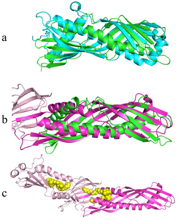
Figure 2.

Examples of structurally similar proteins to Der p 7. Der p 7 is shown in green in all panels. Panel a is the best alignment with JHBP (cyan). Panel b shows the best alignment of Der p 7 with the N terminal domain of BPI shaded magenta, while the C terminal domain of BPI is shaded pale pink. Panel c shows BPI with two bound DSPC molecules (yellow spheres).
