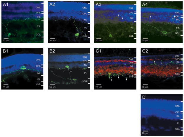
Fig. 2.
Melanopsin-positive RGCs in cross sections of rat retina. All are single (non-stacked) confocal images from 30-μm retinal sections, with a depth of field of only a few micrometers. Melanopsin fluorescent immunolabeling is in green and nuclear counterstaining is in blue. In all images, the brightness of the nuclear staining has been reduced to show the processes in or around the INL; as a result, the nuclear staining in the GCL is quite faint. ONL, outer nuclear layer; OPL, outer plexiform layer; INL, inner nuclear layer; IPL, inner plexiform layer; GCL, ganglion cell layer. (A1 to A4) Nondisplaced RGCs. Arrows in (A3) and (A4) indicate processes in the INL; the contrast and brightness of these two images have been enhanced to show these processes. (B1 and B2) Displaced RGCs. The contrast and brightness in (B2) have also been enhanced to show the axon (arrow). (C1 and C2) Double immunostaining of melanopsin and the presynaptic protein synaptophysin (red Cy3 fluorescence). The punctate melanopsin labeling of the processes (see arrows) did not colocalize or juxtapose with synaptophysin labeling. The cell in (C2) is a displaced RGC. (D) Preadsorption of antibody with the peptide-BSA conjugate abolished all immunostaining.

