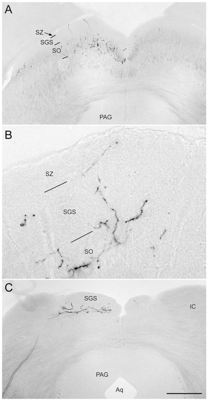
Fig. 10.
Coronal sections illustrating X-gal staining of melanopsin afferents to the superior colliculus. The left eye was enucleated, leaving only right-eye afferents intact. A: Rostral superior colliculus. Fiber labeling is heaviest in the stratum opticum (SO), especially medially and laterally, but some fibers invade the stratum griseum superficiale (SGS) and stratum zonale (SZ). B: Higher magnification view of left colliculus in a similar section from a different brain. C: Caudal pole of superior colliculus, from the same brain as shown in A. In A and C, a few melanopsin afferents from the ipsilateral eye are apparent in the right colliculus. For abbreviations, see list. Scale bar in C = 500 μm in A,C and 200 μm in B.

