Fig. 5.
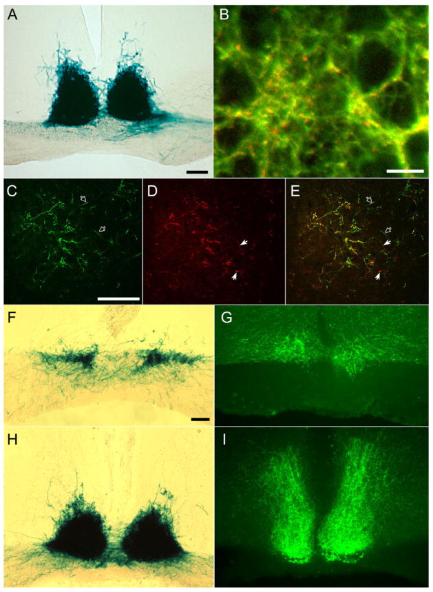
Axonal terminations of melanopsin ganglion cells in the suprachiasmatic nucleus of the hypothalamus. A: X-gal staining of melanopsin afferents to the SCN after right monocular enucleation. Although most labeled fibers in the contralateral (left) optic tract have degenerated, leaving only weak, granular labeling, labeling of the SCN is bilaterally symmetric. B: Confocal image of SCN showing double immunostaining for all contralateral retinal inputs (red; anti-cholera toxin B [CTB] immunofluorescence) and for all bilateral melanopsin RGC inputs (green; anti-β-galactosidase immunofluorescence). Most red, CTB-labeled afferents exhibit green anti-β-galactosidase labeling also, giving them a yellow appearance in the image, but a few contain little if any green labeling and presumably arise from non-melanopsin RGCs. Green axons lacking red immunofluorescence are presumably melanopsin afferents from the ipsilateral eye, which was not injected with CTB. Large circular dark profiles are unlabeled somata of SCN neurons. Projected stack of seven 1-μm confocal optical sections. C–E: Confocal image (optic section thickness 1 μm) grazing the caudal margin of the SCN from the same brain as in B and labeled by the same double-immunofluorescence method. Reduced afferent density permits individual axons to be resolved. Filled arrows indicate crossed retinal afferents (red) that are β-galactosidase negative (no green). Open arrows denote presumptive ipsilateral afferents from melanopsin RGCs (green, no red). F–I: Comparison of the distribution within the SCN and vSPZ of axons of melanopsin ganglion cells (X-gal staining in F and H) with those of VIP-immunoreactive processes in serially adjacent sections. F and G are from the rostral pole of the SCN; H and I are from the mid-caudal SCN. Wisps of fiber labeling dorsal to the main SCN field in H lie within the vSPZ. Scale bar = 100 μm in A, C (applies to C–E), and F (applies to F–I); 10 μm in B.

