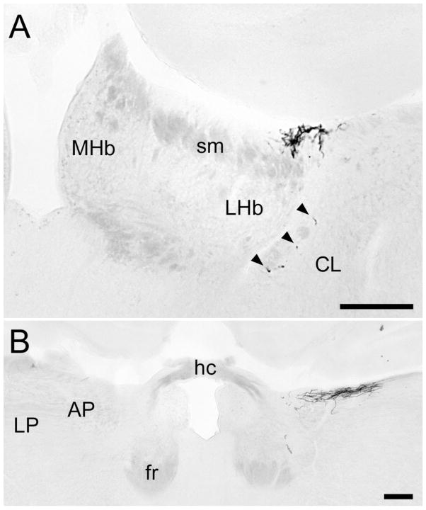
Fig. 8.
X-gal-stained coronal sections illustrating inputs to the region of the habenula and posterior limitans nucleus. A: Mid-habenular level; midline is at the left edge of the image. The main terminal field lies at dorsolateral margin of the stria medullaris and lateral habenula, with scattered fibers (arrowheads) within the fiber capsule bounding the habenular complex ventrolaterally. B: The caudal end of the terminal field, at the level of the habenular commissure. The plexus of labeled fibers on the right lies at the dorsal surface near the boundary of the midbrain (represented by the anterior pretectal nucleus) and the thalamus (represented by the lateral posterior nucleus), in a region included within the posterior limitans nucleus in other rodents. The right eye was enucleated in this animal, leading to nearly complete loss of labeled axons on the left. For abbreviations, see list. Scale bar = 200 μm in A,B.

