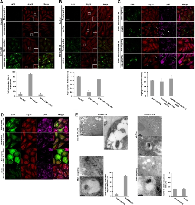
Figure 6.
LC3B and GATE-16 act distinctively in autophagosomal biogenesis. (A, B) Control HeLa cells and HeLa cells stably expressing GFP-LC3B (A) or GFP-GATE-16 (B) were transfected with the reciprocal siRNA pools using DharmaFect reagent. After 72 h interval, the cells were incubated for 2 h in EBSS, subjected to immunostaining with anti-Atg16 after fixation, and analysed by confocal microscopy. Quantification of cells containing Atg16 structures larger than 2 μm (A) or Atg16-labelled puncta structures (B) from three independent experiments in comparison to control and to G to A mutants (D) is presented at the lower panel. Scale bar: 20 μm. (C) HeLa cells stably expressing silent GFP-LC3B or GFP-GATE-16 were transfected with their siRNA pools using DharmaFect reagent. After 72-h interval, the cells were incubated for 2 h in EBSS medium and subjected to immunostaining with anti-Atg16 and anti-p62 antibodies followed by fixation. The cells were analysed by confocal microscopy. Arrows represent cells, which do not express GFP proteins whereas arrowheads represent cells, which express GFP-proteins. Quantification of Atg16-labelled puncta structures from three independent experiments is presented at the right panel. Scale bar: 20 μm. (D) HeLa cells stably expressing GFP-LC3BG120A or GFP-GATE-16G116A were transfected with either pool of GABARAP/GATE-16 siRNAs or LC3 siRNAs, respectively, and a non-targeting siRNA. The cells were starved for 2 h, fixed, and immunostained with anti-Atg16 and anti-p62 antibodies. Quantification is presented in (A, B) at the lower panels. Scale bar: 20 μm. (E) HeLa cells stably expressing GFP-LC3B (left panel) or GFP-GATE-16 (right panel) were transfected with the reciprocal siRNA pool or with non-targeting siRNA and starved for 2 h in EBSS medium. The cells were fixed and their cryo sections were immuno-labelled using anti-GFP antibodies and analysed by TEM as described in ‘Materials and methods'. Quantification of cells containing Atg16 structures larger than 2 μm (left panel) or the size of Atg16-labelled structures (right panel) is presented.

