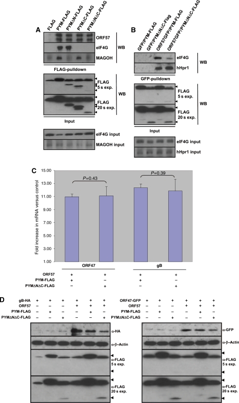
Figure 6a-d.
An ORF57-specific dominant negative PYM mutant prevents ORF57 association with the 48S pre-initiation complex and perturbs ORF57-mediated intronless KSHV mRNA translation. (A) 293T cells were transfected with pORF57GFP in addition to either pFLAG, pPYM-FLAG, pPYMΔN19-54-FLAG, pPYMΔC53-FLAG and pPYMΔNΔC-FLAG, and immunoprecipitations were performed using FLAG-affinity beads. Western blot analysis was then performed using the indicated antibodies to detect the precipitated proteins. To detect the expression of each PYM-FLAG protein (indicated by the solid triangles), total cell lysate was analysed by western blotting using an FLAG-specific antibody. Two different autoradiograph exposures (exp.) are shown (5 and 20 s), as PYMΔNΔC is fused to a single FLAG epitope (as opposed three for the other fusion proteins) and therefore generates a weaker signal. Loading controls for each co-immunoprecipitation are also shown. (B) 293T cells were transfected with either pGFP or pORF57GFP in addition to either pPYM-FLAG or PYMΔNΔC-FLAG and an immunoprecipitation performed using a GFP-specific antibody. Western blot analysis was then performed using the indicated antibodies to detect precipitated proteins. Loading controls for each co-immunoprecipitation are also shown. (C) 293T cells were triple transfected with either pORF47GFP or pgB-HA, in the presence or absence of pCherry or pORF57Cherry and either pPYM-FLAG or pPYMΔNΔC-FLAG. At 24 h post-transfection, cytoplasmic RNA was isolated from each sample and 1 μg of RNA was used to generate cDNA using reverse transcriptase. A measure of 10 ng of cDNA was then used in qRT–PCR reactions. Fold increase was determined by ΔΔcT and statistical significance by a non-paired t-test. All data are representative of three independent experiments and presented as fold increase versus pCherry-transfected controls. (D) 293T cells were triple transfected with either pCherry or pORF57Cherry in addition to either pFLAG, pPYM-FLAG or pPYMΔNΔC-FLAG and either pORF47GFP or pgB-HA. At 24 h post-transfection, western blot analysis was performed using 25 μg of total protein extract and the indicated antibodies to detect protein expression levels. To detect the expression of PYM-FLAG and PYMΔNΔC-FLAG, total cell lysate was analysed by western blotting using a FLAG-specific antibody. Two different autoradiograph exposures (exp.) are shown (5 and 20 s) as PYMΔNΔC is fused to a single FLAG epitope (as opposed three for the other fusion proteins) and therefore generates a weaker signal.

