
Figure 4. Transcriptional suppression of IIS genes in age-1(mg44).
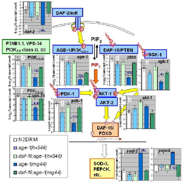
Transcript levels were assayed by real-time polymerase chain reaction (RT-PCR). Expression histograms are shown superimposed on a schematic diagram of IIS. Yellow arrows show protein phosphorylations (circled P's); orange arrows indicate binding of phosphatidylinositol 3,4,5-triphosphate (PIP3, “structural” symbols). Within each histogram, transcript mean ± SEM (steady-state) is shown on a log(2) scale, comparing wild-type to 4 age-1 mutant groups and to dauer larvae. For each group, fold changes are shown (e.g., “3×”), of age-1 (mg44)-F2 relative to N2drm. Post-gravid age-1(mg44) F1 homozygotes were at adult day 8–9; F2 homozygotes at day 10; N2drm, age1(hx546), and daf-16(mu86); age-1(mg44) double mutants, were all post-gravid adults at adult day 6; and N2drm dauer larvae 1 day after reaching 98% SDS-resistance. Transcript levels are means of 3 independent biological replicates, normalized to the mean of three control gene (β-actin, T08G5.3, and Y71D11.3) that did not differ significantly among strains.