
Fig. 4.
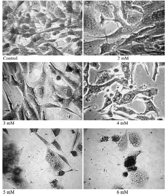
Induction of vacuoles in cocaine treated cells. Glial cells were treated with 2–6 mM cocaine continuously for 24 h in the incubator. Monolayer cells were fixed and stained with 0.1% Crystal violet dye, and photographed using inverted phase contrast IX-70 Olympus microscope with 40X objective