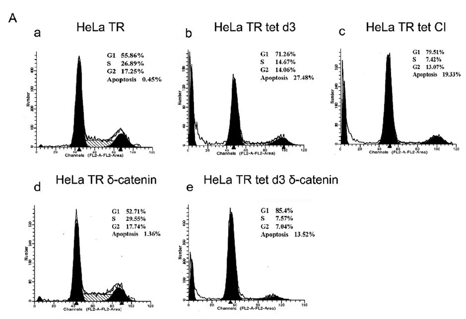
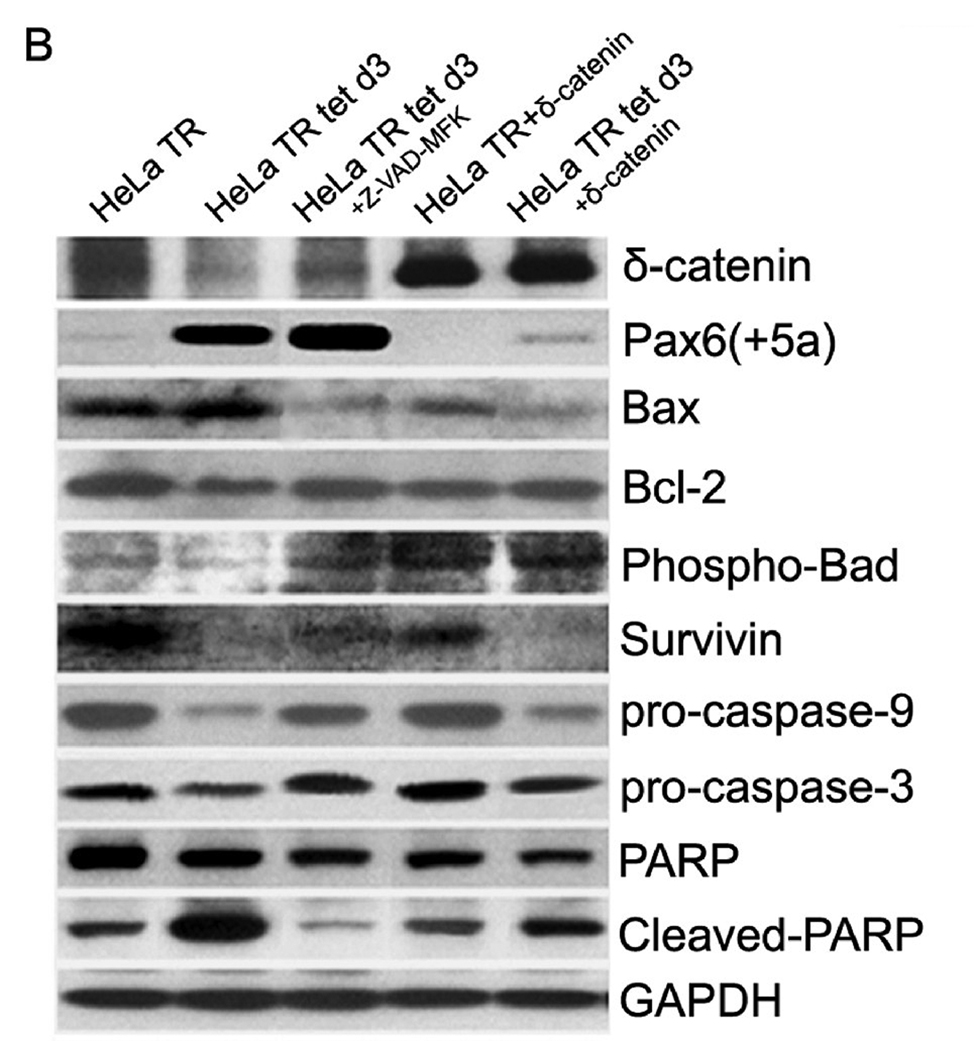
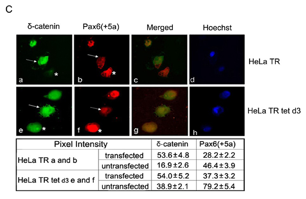
Fig. 6. Mitotic and apoptotic changes in HeLa Tat-TetR-Pax6 cell line.
A. Cell cycle kinetics analysis. HeLa Tat-TetR-Pax6 cells after doxycycline induction is treated respectively with caspase inhibitor (Z-VAD-FMK) or transfected with δ-catenin. At day3, cells in each group are collected and analyzed by flow cytometry with propidium iodide (PI) staining. a: HeLa Tat-TetR-Pax6 cells without doxycycline induction (HeLa TR); b: HeLa Tat-TetR-Pax6 cell 3 days after doxycycline induction (HeLa TR tet d3); c: HeLa Tat-TetR-Pax6 cell with Pax6(+5a) overexpression in the presence of Z-VAD-FMK (HeLa TR tet CI); d: Overexpression of δ-catenin in HeLa Tat-TetR-Pax6 cells without Pax6(+5a) overexpression (HeLa TR δ-catenin); e: Overexpression of δ-catenin in HeLa Tat-TetR-Pax6 cells with Pax6(+5a) overexpression (HeLa TR tet δ-catenin). B. The effects of Pax6 and δ-catenin expression on apoptotic protein expression. Western blots show protein expression profiles of Pax6(+5a), δ-catenin, PARP, cleaved-PARP, procaspase-9, procaspase-3, Bax, Bcl-2, Phospho-Bad and survivin. Note that δ-catenin expression in HeLa TR tet d3 cells is reduced with high Pax6(+5a) expression when compared to that in HeLa TR cells. Blocking caspase activation using caspase inhibitor (HeLa TR tet d3+Z-VAD-FMK) reduces the inhibitory effects of high Pax6(+5a) expression on δ-catenin expression. Pax6(+5a) and δ-catenin expression is both increased when compared to that of no-caspase inhibitor treatment. On the other hand, overexpression of δ-catenin (HeLa TR tet d3+δ-catenin) also reduces Pax6(+5a) expression and the apoptotic protein expression induced by Pax6(+5a) overexpression. The results show that overexpression of δ-catenin reduces Pax6(+5a) expression in HeLa Tat-TetR-Pax6 cells, which provides a negative feedback between Pax6 and δ-catenin expression. C. Double immunofluorescent light microscopy showing the effects of δ-catenin overexpression on Pax6(+5a) expression. a–d: Pax6(+5a) (red) and δ-catenin (green) expression in HeLa TR cells; c: Merged image of a and b; d: Nuclei staining. e–h: Pax6(+5a) (red) and δ-catenin (green) expression in HeLa tet d3 cells. g: Merged image of e and f; h: Nuclei staining. Note: arrows indicate δ-catenin transfected cells; asterisks indicate untransfected cells. The arrows show that the expression of Pax6(+5a) (red) is reduced in cells transfected with δ-catenin (green). The table in C shows the morphometric density of pixel values in cells stained by immunofluorescence light microscopy. Bar: 20 µm.


