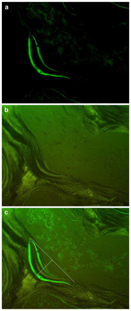
Fig. 1.
Photomicrographs of a typical basic multicellular unit (BMU) for which analysis of resorption parameters was conducted. a BMU viewed with epifluorescence to identify calcein labeling which indicates active bone remodeling. b BMU viewed with polarized light to identify the cement line which indicates the limit to erosion of the remodeling unit. c Overlay of the two images used for measurement of BMU morphology along with lines depicting how the surface of the cavity was estimated for assessment of resorption cavity width (Rs.Wi) and depth (Rs.De). Resorption cavity area (Rs.Ar) was defined as the space within the entire cavity, Images are shown at original magnification ×250

