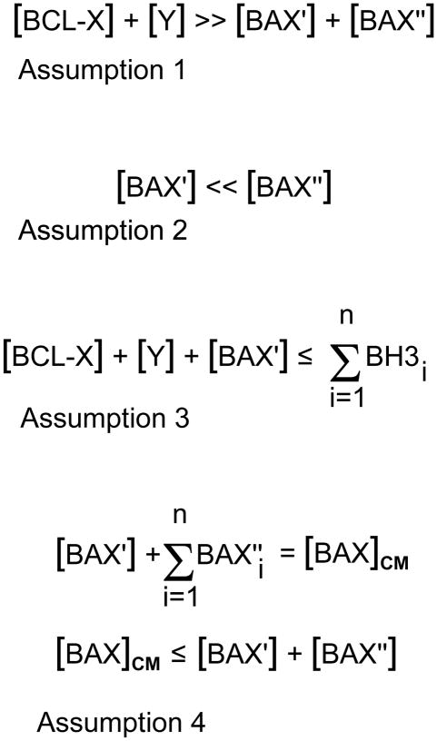
Fig. 4.
Mathematical assumptions governing variations in the rheostat model. In this series of equations, important aspects of the different levels of members of the Bcl2 gene family proteins are considered for their roles in blocking apoptosis and/or in the activation of the intrinsic cell death program. Variables are given as either cellular concentrations (written in square brackets) or as absolute numbers of molecules, in which each variable is written in summation form. The variables are:
BCL-X = the principal anti-apoptotic protein expressed in retinal ganglion cells, Y = other molecules of the Bcl2 gene family with anti-apoptotic properties. This may include, but not be limited to, molecules such as BCL2, MCL1, BCL-W, etc.
BAX′ = molecules of BAX that are initially converted from a globular soluble structure to become inserted in the mitochondrial outer membrane. For the purposes of this argument, this is considered the primary BAX activating event, and probably requires interaction with one or more members of the BH3-only peptide family.
BAX″ = all other latent BAX molecules that are present in the cytosol, but are not activated by BH3-only peptide interactions. Instead, these molecules become the pool of BAX proteins that are secondarily activated after the unfolding and insertion of BAX′ molecules.
BAXCM = the critical mass of BAX molecules that must be assembled as oligomers in the mitochondrial membrane. This is a hypothetical variable and makes the assumption that functional destabilization of the mitochondrial membrane requires a specific density of BAX proteins to assemble.
BH3 = BH3-only peptides that become activated during the course of apoptosis of ganglion cell somas.
Assumption 1. This equation summarizes the probable association of BAX molecules and anti-apoptotic molecules present in living, healthy, ganglion cells. Semi-quantitative western blots to measure protein levels, and quantification of transcripts using real-time PCR suggest that just BCL-X alone is several fold more abundant than BAX levels in ganglion cells. Although BAX must become activated in order to execute the cell death program, the excess of antagonistic BCL-X likely prevents “accidental” activation events, which may be one reason why BCL-X is already localized to the surface of organellar membranes.
Assumption 2. This equation summarizes the hypothetical assumption that BAX activation occurs in two stages. The first stage requires only minimal initial activation of BAX (BAX′) by interaction of globular BAX with a BH3-only protein. Once unfolded, BAX′ is able to both insert into the mitochondrial outer membrane, and act as a sink to activate other molecules of globular BAX in the cytosol (BAX″). This event, by itself, is probably not sufficient to elicit cell death, but is likely sufficient to initiate recruitment of other BAX proteins.
Assumption 3. This equation summarizes the critical event leading to the activation of BAX′ molecules, and is essentially the core element of the modern rheostat model. In this model, enough BH3-only proteins must be recruited to inactivate all anti-apoptotic proteins present in a living cell ([BCL-X] + [Y]) as well as still be available to activate BAX′. Equilibrium effects, in which the binding of BH3-only proteins is favored for some Bcl2 family proteins over others, probably requires and excess of BH3-only peptides (n) in order to form partners with all the relevant variables in this equation.
Assumption 4. These two equations summarize the threshold requirements for BAX in order to form functional oligomers in the mitochondrial outer membrane. First, a critical mass of activated BAX is required for oligomer formation. The critical mass is composed of BAX′ molecules, and some portion (n) of BAX″ molecules. It is likely, however, that cells generally contain enough latent BAX molecules to achieve BAXCM, and cells with large excesses of BAX do not exhibit a greater cell death response once critical mass is established. Cells with BAX content below the critical mass threshold are resistant to apoptosis, however. In this model, I assume that [BAX′] ≪ [BAXCM], therefore, subthreshold levels of BAX probably affect the efficiency of secondary recruitment of BAX″ molecules to the mitochondria, thus delaying the formation of BAXCM. Evidence for this is the very slow activation of apoptosis over several months in Bax+/− retinal ganglion cells that are initially resistant to optic nerve crush (Semaan et al., 2010).

