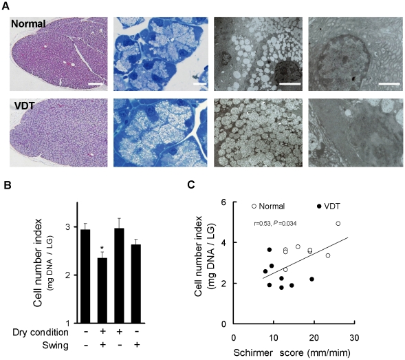
Figure 3. Rat VDT user model causes alterations in lacrimal gland morphology.
(A) Left: H & E staining. Left center: Toluidine blue staining. Right center and right: Electron microscopic analysis of acinar cells. Images showing expanded aciner cells accompanied by accumulated enlarged secretory vesicle in the cytoplasm (center), decresed endoplasmic reticulum and increase in the nuclei with dark neucleoplasm (Right) of LG on day 10. Scale bars: Left = 200 µm; Left and right center = 10 µm; Right = 4 µm. (B) Changes in total cell number of LG. Changes of LG cell number were measured 10 days after treatment with or without swing or dry condition. Quantification of LG number was calculated by deoxyribonucleic acid content of the LG. Data represent the mean ± SEM for 8 to 16 eyes. * P<0.05 versus the without swing and dry condition. (C) Correlation between tear production and LG cell number. Pearsons correlation coefficient testing was r = 0.53 (P = 0.034, n = 16).

