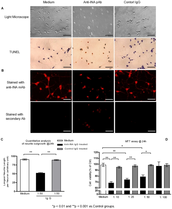
Figure 6. The anti-INA pAb inhibit the growth of neurites and result in neuron apoptosis in vitro.
(A) Primary rat embryo neurons were co-cultured with medium, sterile anti-INA pAb (1∶50), or control IgG for 96 hrs. The neurite outgrowth and cell viability was observed by light microscopy. Apoptotic cells were detected by TUNEL assay. (B) A monoclonal antibody (Mab 5224) against INA was used for displaying INA location. The TRITC goat anti-rabbit secondary antibody staining was to demonstrate the internalization of co-cultured Abs during the neuron cell growth. (Bar = 25 µm) (C) Quantitative measurement of cultured neurons axonal length at 24 hrs. (D) Viability of primary cultured neurons subject to indicated concentrations of anti-INA pAb or control IgG after 24 h. The percentage of cell survival normalized to control cells was applied in MTT assay. Data are represented as mean±SEM of three independent experiments.

