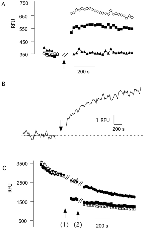
Figure 7. sPB1-F2 generates Ca2+ and anion fluxes into liposomes.
(A) Fluorescence of liposomes with Ca2+ sensitive dye Fluo3 was recorded before and after adding (at arrow) ionophore Valinomycin (triangle), sPB1-F2pr8 alone (filled squares) or together with Valinomycin (open squares). Peptide and ionophore were added during the time gap of ca. 1 min indicated in the graph. The presence of the peptide results in an increase in fluorescence indicating an influx of Ca2+ into the liposomes. The ionophore enhances Ca2+ influx because it prevents building up of a charge, which hinders net Ca2+ influx. (B) Fluorescence of liposomes filled with Ca2+ sensitive dye Fluo-3 before and after addition (at arrow) of 1 µM peptide to incubation medium. The truncated peptide sPB1-F2pr8 50–87 results in a fast rise in Fluo3 fluorescence. (C) Fluorescence of liposomes filled with anion sensitive dye lucigenin was measured before and after adding of anion specific ionophore TBT (filled squares, added at arrow 1), sPB1-F2pr8 (open triangle, arrow 2). The control was left untreated (filled circles); the stepwise drop of the control signal is due to an unspecific drift of the signal. Both ionophore and sPB1-F2pr8 generate a strong quenching of the lucigenin fluorescence well beyond the control indicating an influx of anions. Peptide and ionophore were added during the time gap of ca. 1 min indicated in the graph.

