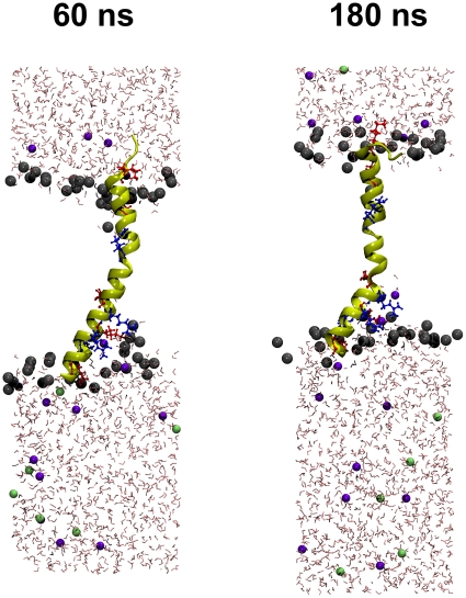
Figure 10. Snapshots of the simulation system after removal of the center-of-mass constraint (set to 0 ns).
The protein is shown in cartoon representation with explicit depiction of positively charged residues (arginine: blue, lysine: red). Lipid molecules have been removed except for the head groups that are depicted as grey spheres. Potassium ions are shown in green, chloride ions in blue. The c-terminus is located on the bottom side.

