
Figure 2. Spontaneous Th17-dominated skin inflammation in 8 week-old ft/ft mice.
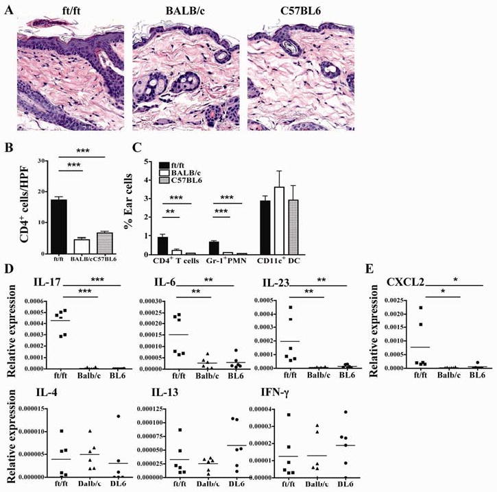
(A) Representative photomicrographs of H&E sections. (B) Numbers of CD4+ cells/HPF (400×). (C) FACS analysis of CD3+CD4+ cells and CD11b+Gr-1+ neutrophils in ear skin. (D, E) Cytokine (D) and CXCL2 (E) mRNA expression in the skin. *p<0.05, **p<0.01, ***p<0.001.