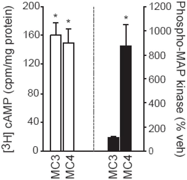
Figure 1.
cAMP and MAP kinase signaling through the melanocortin receptors. COS-1 cells were transfected with MC3 or MC4 receptor and treated with MTII. Formation of cAMP is shown on the left (white bars) and activated (phosphorylated) p44/42MAP kinase is shown on the right (black bars). Asterisks are used to indicate differences from cells in the same transfection condition that were treated with vehicle. Based on Daniels et al. [29].

