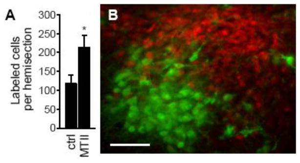
Figure 2.
Melanocortin-induced p44/42MAP kinase activation in rat PVN. (A) The number of cells immunohistochemically labeled for phosphorylated p44/42MAP kinase in brains from rats treated with vehicle or MTII is shown as the mean (±SEM) cells per PVN hemisection). An asterisk indicates p<0.05. (B) Double-labeling for phosphorylated p44/42MAP kinase (red) and oxytocin (green) in the brain of a rat injected with MTII. The scale bar is 50 μm. Data are from Daniels et al. [29].

