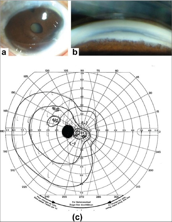
Figure 1.

Photographs of the anterior segment of a 42-year-old woman with Axenfeld- Rieger syndrome. (a) Anterior segment photographs of the left eye of the proband at the age of 42 years showing microcornea, iris hypoplasia (arrows), and posterior embryotoxon (arrowheads). Deposits are seen at the corneal limbus which may reflect aging changes. Nuclear cataracts were also present. (b) Gonioscopic images of the same patient showing an abnormal angle and iridocorneal tissue adhesions traversing the anterior chamber. (c) Goldmann kinetic perimetric fields of the patient at the age of 42 years
