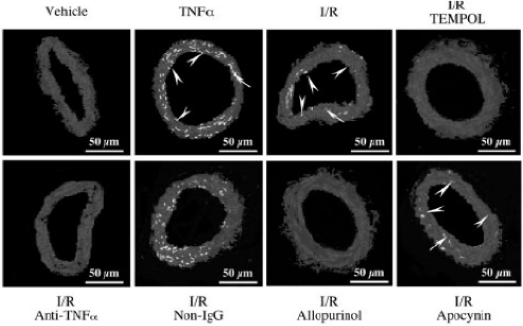
Fig 1.
DHE-fluorescence imaging of O2 in coronary arterioles . DHE fluorescence was markedly elevated in both endothelial (arrow head) and vascular smooth muscle (arrow) cells of arteriolar sections after TNF-α treatment (1 ng/mL, 60 minutes, n=4) and I/R (n=4) compared with vehicle (sham). Apocynin and nonimmune IgG did not, but TEMPOL, allopurinol, and anti-TNF-α, markedly reduced the fluorescent signals (n=4). (Adapted from Figure 1 in Zhang, C et al. [129].

