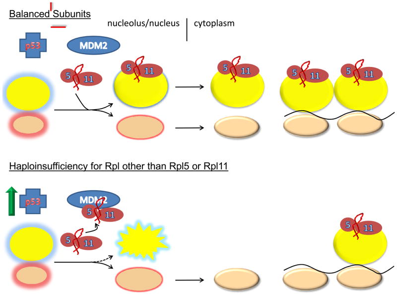
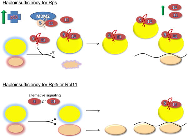
Figure 2. Models of ribosomal stress.
2a Balanced subunits, schematic represents the normal state with the balanced production of 40S and 60S subunits. Immature subunits are shown in the nucleolus/nucleus with colored glows. Mature subunits in the cytoplasm are either free or engaged in translation as polysomes on the black line representing mRNA. Under these conditions there is insufficient free ternary complexes of Rpl5, Rpl11 and 5S rRNA to bind MDM2 and block its interaction with p53. Thus, p53 levels are relatively low. 2b shows the situation when essential large subunit ribosomal proteins other than Rpl5 and Rpl11 are depleted. Under these conditions large subunit assembly is disrupted and the Rpl5/Rpl11/5S rRNA ternary complex is free to interact with MDM2. Interaction of the ternary complex with MDM2 interferes with the ability of MDM2 to interact with p53 resulting in p53 stabilization. The third panel shows the situation when essential 40S ribosomal subunit proteins are depleted. Under these conditions it is shown that TOP mRNAs including Rpl11 are upregulated. Excess Rpl11, presumably in complex with Rpl5 and 5S rRNA is then available for interaction with MDM2 resulting in p53 activation. In this manner, signaling through large subunit ribosomal components is possible even though it is the small subunit, whose assembly is disrupted. Finally, the last panel shows the situation when either Rpl5 or Rpl11 is depleted. Here accumulation of the ternary complex signaling module is lost creating a situation where alternative means of signaling ribosome stress must be employed. These alternative signaling mechanisms may be less effective and give rise to distinct and more severe clinical phenotypes.


