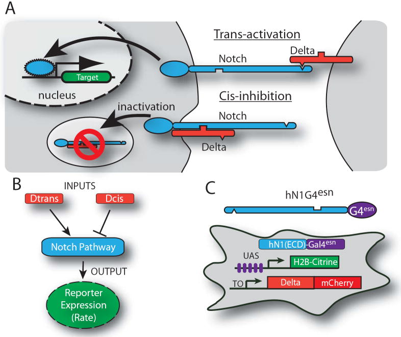
Figure 1. System for analyzing signal integration in the Notch-Delta pathway.
(A) Notch (blue) and Delta (red) interactions are indicated schematically. (B) Notch activity integrates cis- and trans-Delta. (C) CHO-K1 cell line for analyzing Notch activity. (C) The hN1G4esn cell line stably incorporates a variant of hNotch1 in which the activator Gal4esn replaces Notch ICD. This cell line also contains genes for Histone 2B (H2B)-Citrine (YFP) reporter controlled by a UAS promoter, a Tet-inducible Delta-mCherry fusion protein, and a constitutively expressed H2B-Cerulean (CFP) for image segmentation (not shown). A similar cell line expressing full length hNotch1 (hN1 cell line) was also analyzed (Figs. S1, S2). These cells exhibit no detectable endogenous Notch or Delta activities. Notch-Delta interactions are indicated schematically and do not represent molecular interaction mechanisms11.

