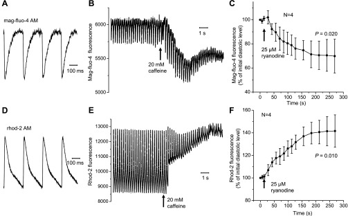
Fig. 1.
Changes in fluorescence from the sarcoplasmic reticulum (SR) lumen (A–C) and cytosol (D–F) measured with the Ca2+-sensitive dyes rhod-2 AM and mag-fluo-4 AM, respectively. Representative traces of luminal (A) and cytosolic (D) Ca2+ transients are shown. Diastolic (resting) levels of mag-fluo-4 (B) and rhod-2 (E) fluorescence changed in different directions in response to the application of caffeine. An analogous effect was observed in the presence of ryanodine. Changes in mag-fluo-4 (C) and rhod-2 (F) fluorescence reflect the release of Ca2+ from the SR and accumulation of Ca2+ in the cytosol, respectively. Data are means ± SD. All measurements were conducted at 4 Hz and 37°C.

