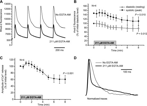
Fig. 3.
Effect of 4-min perfusion with Tyrode solution containing 211 μM EGTA AM on Ca2+ dynamics in the cytosol measured with rhod-2 AM (A–C). Measurements were conducted at 37°C at a heart rate of 4 Hz. A: representative traces obtained before (thin line) and 4 min after (thick line) perfusion with EGTA AM. B: changes in the diastolic (resting) and systolic (peak of transient) levels of rhod-2 fluorescence during 4-min treatment with EGTA AM. C: reduction in the amplitude of Ca2+ transients after the addition of EGTA AM. The period of treatment with EGTA AM is indicated by the shaded bar. Data are means ± SD; N is the number of independent experiments (hearts). P values are the results of one-way ANOVA (initial vs. final levels of the parameters). D: typical traces of fluorescence from mouse hearts loaded with rhod-2 AM in the absence (thin line) and presence (thick line) of 211 μM EGTA AM (37°C, 2 Hz). Traces were normalized to the maximum amplitude of the release for the comparison of the kinetics.

