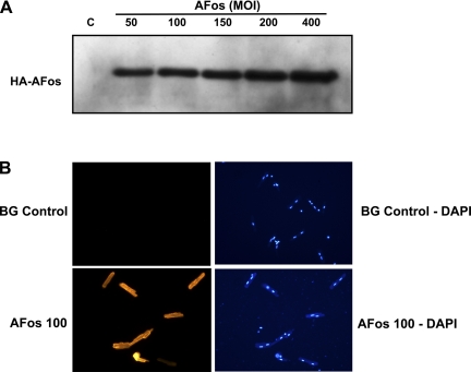
Fig. 1.
Determination of AFos expression in the adult rat ventricular myocytes. A: myocytes were infected with AFos in increasing MOI (50–400 MOI) for Western blot analysis and compared with β-galactosidase (β-Gal; BG) control (C) [100 multiplicity of infection (MOI)]. Anti-hemagglutinin (HA) antibody diluted 1:1,000 shows increasing signal corresponding to increasing expression of AFos. B: myocytes were infected with β-Gal (100 MOI) vs. AFos in increasing MOI. 4,6-Diamino-2-phenolindole (DAPI) stain shows viable myocytes with binuclear expression pattern with corresponding immunofluorescence in rod-shaped myocytes.

