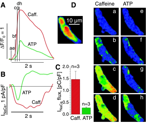
Fig. 3.
Caffeine- and ATP-activated Ca2+ transients and inward currents in juvenile cardiomyocytes. A: superimposed cellular Ca2+ transients (ΔF/F0) upon extracellular application of 10 mM caffeine (Caff; red) and 100 μM ATP (green) for the voltage-clamped cell shown in the inset and in D. B: membrane current recorded at −70 mV during 2-s exposures to caffeine (red) and ATP (green). The duration of the solution exchange is indicated by the 2-s time bars in A and B. C: average values for Na+-Ca2+ current (INaCa)-flux during exposure to caffeine and ATP are based on the integral of the measured membrane current during its negative excursion (B). D: normalized images of Ca2+-dependent fluorescence before activation (a and e) and during exposure to caffeine (b–d) or ATP (f–h) at the times indicated in B. Voltage-clamped juvenile cells are same color scale as Fig. 1.

