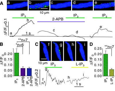
Fig. 5.
IP3-gated Ca2+ signals in juvenile, permeabilized cardiomyocytes are reversibly suppressed by the IP3-gated Ca2+ release channels (IP3R) antagonist 2-aminoethoxydiphenylborate (2-APB; A and B) and are triggered only weakly by L-IP3 (AB). A and C: normalized fluorescence images that were recorded in representative cardiomyocytes at the times indicated next to the tracings of cellular Ca2+ transients, i.e., before interventions (a, f, and h), during activation with 20 μM IP3 alone (b and g), and when IP3 was applied after 3 min incubation with 2 μM 2-APB (c), after 3 min of washout (e), or as the biologically inactive stereoisomer L-IP3 (i). B: average amplitude of fluorescence signals (ΔF/F0) evoked by IP3 (green) in cells that were then incubated with 2-APB (violet) and again challenged with IP3 (turquoise). D compares the average amplitudes of the cellular Ca2+ transients in another set of cells that were exposed sequentially to IP3 (green) and L-IP3 (yellow; permeabilized cells, paired t-test). *P ≤ 0.05; **P ≤ 0.01.

