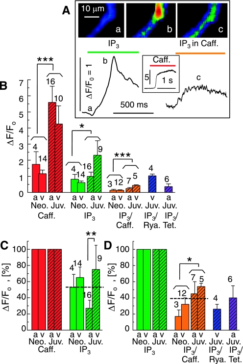
Fig. 6.
Contribution of ryanodine receptors (RyR)-gated Ca2+ stores to the IP3-triggered Ca2+ releases. A: cellular Ca2+ transients (ΔF/F0) in a permeabilized juvenile cardiomyocyte upon extracellular application of 20 μM IP3 (left), 10 mM caffeine (inset), and IP3 after 3 min incubation in caffeine (right). Selected frames (a-c) show normalized fluorescence images at the times indicated. B: comparison of the average Ca2+ transients (ΔF/F0) in atrial (a) and ventricular (v) cardiomyocytes from neonatal (Neo; open bars) and juvenile (Juv; hatched bars) rats. The Ca2+ transients were evoked by 10 mM caffeine (red) or 20 μM IP3, either by itself (green) or after 3 to 4 min incubation with 10 mM caffeine (orange), 40 μM ryanodine (blue), or 1 mM tetracaine (purple). C: IP3-activated Ca2+ transients normalized relative to the caffeine-induced transients measured in the same cells. D: suppression of IP3-activated Ca2+ signals by pretreatment with caffeine, ryanodine, and tetracaine. Permeabilized cells are same color scale as Fig. 1; n values are indicated at each bar. *P ≤ 0.05; **P ≤ 0.01; ***P ≤ 0.001.

