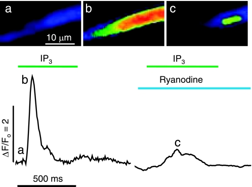
Fig. 9.
IP3-mediated perinuclear Ca2+ release in a permeabilized juvenile cardiomyocyte. Representative cellular Ca2+ transients (ΔF/F0) and fluorescence images (a–c) evoked by extracellular application of 20 μM IP3 before (left) and after (right) 5-min incubation in 40 μM ryanodine. The normalized fluorescence images were recorded at 240 frames/s before activation (a) and at the peak of the IP3-activated responses, which normally invaded the entire cell (b) but were confined to the nuclear region after ryanodine treatment (c).

