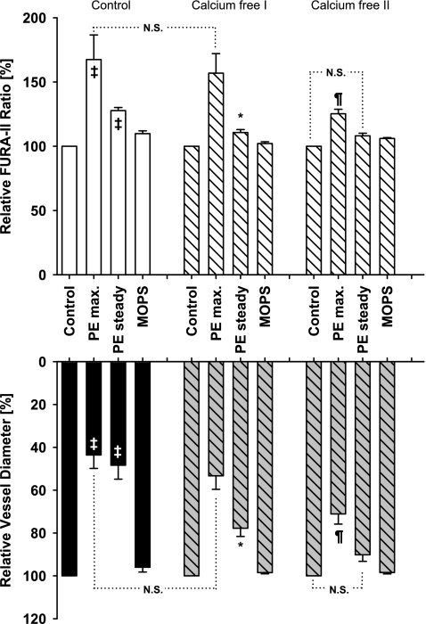
Fig. 4.
Effect of extracellular calcium ion removal on PE-induced constriction. In calcium containing buffer (control), vessels from iPLA2β knockout mice constricted significantly to extraluminal PE (100 μ mol/l) concurrent with a significant increase in calcium levels (‡P < 0.05 from respective controls, ANOVA; n = 7). Calcium removal strongly attenuated steady-state constriction (calcium free I; *P < 0.05, ANOVA), but not initial maximum constriction (NS) or initial calcium response (NS) compared with calcium control consistent with the initial constriction dependent on intracellular calcium release and the steady-state constriction on extracellular calcium influx. A second stimulation with PE (calcium free II) greatly diminished the initial maximum constriction (¶) and calcium response (¶), indicating that intracellular calcium stores have been depleted (calcium free 2, right). Furthermore, the vessel dilated within seconds with the diameter and the calcium level no longer different from calcium free II control (NS).

