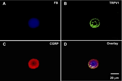
Fig. 2.
Colocalization of transient receptor potential vanilloid 1 (TRPV1) and CGRP on kidney projecting sensory neurons using fluorescence microscopy. A: kidney projecting sensory neuron labeled by FB (blue). B: TRPV1- immunostaining (green) on the same neuron. C: CGRP-immunostaining (red) on the same neuron. D: overlay picture combining A, B, and C.

