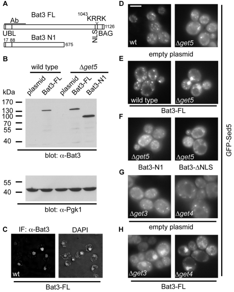
Fig. 6.
Bat3 relocalises GFP-Sed5 in S. cerevisiae GET mutants. (A) Outline of full-length Bat3 (isoform 2), and the N-terminal fragment used in this study. The locations of the ubiquitin-like domain, NLS and BAG domains and the antibody-binding region are indicated. In the ΔNLS mutant, the KRRK motif shown is altered to KRSL to disrupt nuclear targeting of Bat3 (Manchen and Hubberstey, 2001). (B) Immunoblot showing Bat3 and phosphoglycerate kinase 1 (Pgk1) levels in wild-type or Δmdy2 (Δget5)-transformed S. cerevisiae cells. (C) Subcellular localisation of full-length Bat3 expressed in wild-type S. cerevisiae and DAPI staining of nuclei visualised by immunofluorescence microscopy. (D-H) The effect of Bat3 expression upon the subcellular localisation of GFP-Sed5 was determined by live-cell imaging in wild-type, Δget3, Δget4 or Δmdy2 (Δget5) cells, as indicated. Full-length Bat3, an N-terminal fragment or the ΔNLS mutant, were used as indicated. See also supplementary material Fig. S2C for fixed and immunostained cells of the same genotype demonstrating co-localisation of Bat3 and GFP-Sed5 immunoreactivity with DAPI staining of the nucleus in Δmdy2 (Δget5) cells. Scale bar: 5 μm.

