Fig. 4.
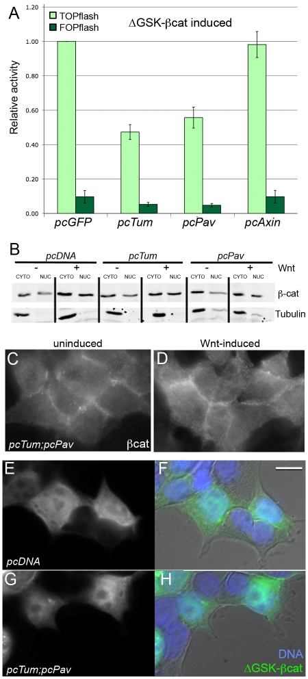
Pav and Tum do not alter β-cat stability or nuclear translocation. (A) TOPflash activity induced by the constitutively-active ΔGSK-βcat mutant is repressed by Tum (P=0.003) and Pav (P=0.009). Axin, a destruction complex component, was not able to repress ΔGSK-βcat (P>0.76). Results are means ± s.d. (B) Immunoblots show that βcat levels in the nuclear fraction of HEK293T cells increased in response to Wnt stimulation, as shown in the pcDNA3.1 empty vector control lanes. This pattern of Wnt-induced nuclear βcat accumulation is not altered in HEK293T cells transfected with either Tum or Pav. β-tubulin was used as a loading and fractionation control. (C) Uninduced and (D) Wnt-induced HEK293T cells that overexpress Tum and Pav show similar patterns of endogenous βcat distribution, with higher overall levels in the Wnt-induced cells. HEK293T cells transfected with ΔGSK-GFP-βcat and stained for GFP show high uniform βcat levels in the transfected cells (E), which have normal morphology compared with neighboring untransfected cells (F, brightfield image merged with fluorescent images). (G) Co-transfection with Tum and Pav does not alter the distribution of ΔGSK-GFP-βcat compared with the empty vector control, nor does it alter cell morphology (H). Scale bar: 10 μm (C-H).

