Fig. 3.
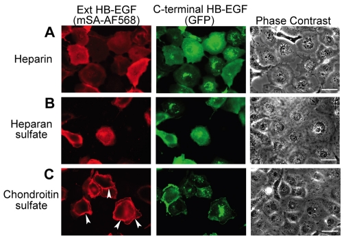
Heparin and heparan sulfate changed the localization of pro-HB-EGF from sites of cell-cell contact to a homogenous distribution over the cell surface. After 24 hours of transfection of COS-7 cells with AP–HB-EGF–GFP, the biotinylated, extracellular acceptor peptide in AP–HB-EGF–GFP was labeled with monovalent streptavidin–Alexa-Fluor-568 (red, left) and imaged alongside the cytoplasmic tail conjugated to EGFP (green, middle), and phase contrast (right). Addition of (A) heparin (100 μg/ml) or (B) heparan sulfate (100 μg/ml) for 4 hours changed the localization of both the extracellular and intracellular domains of AP–HB-EGF–GFP to a diffuse distribution over the cell surface, rather than at sites of cell-cell contact. (C) Addition of chondroitin sulfate (100 μg/ml) for 4 hours had no effect on localization of AP–HB-EGF–GFP. Each row represents the same field. Arrowheads indicate sites of cell-cell contact. Scale bars: 40 μm.

