Fig. 2.
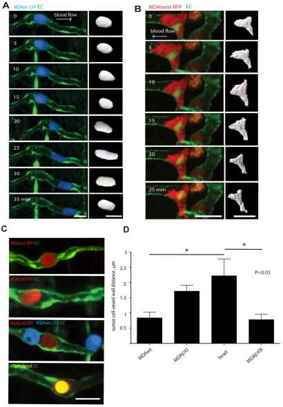
Tumor cells arrest in small vessels then actively migrate along the luminal surface of the vascular endothelium. (A) MDAwt cell expressing CFP and migrating within the ISV lumen for the indicated times (supplementary material Movie 1). Right panel shows 3D isosurface rendering of the tumor cell body for each time point (supplementary material Movie 2). (B) MDAtwist cell expressing RFP and migrating in the lumen of an ISV for the indicated times (supplementary material Movie 4). Right panel shows 3D isosurface rendering of the tumor cell body for each time point (supplementary material Movie 5). (C) Multi-color confocal images of MDAwt and MDAβ1KO cells labeled with CFP or RFP that have arrested in the ISV lumen. Lower panel shows a control 10 μm fluorescent Sepharose bead (yellow) arrested in the ISV. (D) Quantification of distance from tumor cell to vessel wall for MDAwt, MDAβ1KO cells, and Sepharose beads as shown in representative Fig. 2C above. Results are means ± s.e.m. Scale bars: 20 μm.

