
Figure 1.
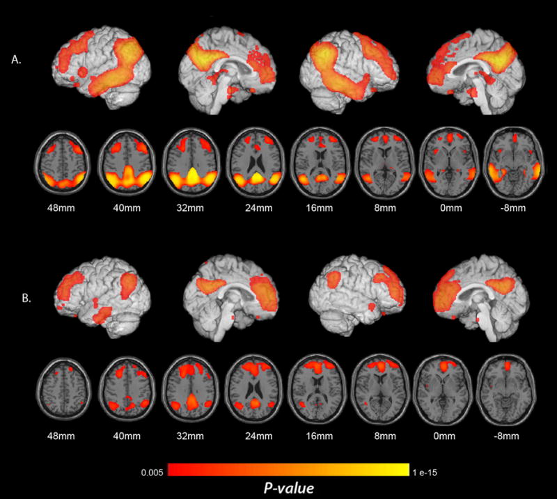
Regions with significantly lower CMRgl in pAD and aMCI patients. (A) Lower CMRgl in pAD patients than in elderly NC. (B) Lower CMRgl in aMCI patients than in elderly NC (p < 0.005, uncorrected for multiple comparisons). Significance levels in these brain maps are uncorrected for multiple comparisons. Findings are projected on to the lateral and medial surfaces of the left and right cerebral hemispheres and are also shown on horizontal sections in relationship to a horizontal section between the anterior and posterior commissures.