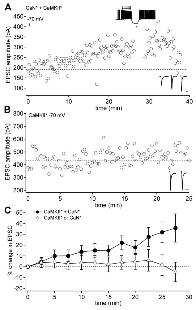
Figure 4.
EPSC amplitudes run up with CaN* and CaMKII* infused together in neurons held at −70 mV. (A) EPSC amplitudes at −70 mV in a neuron infused with both CaN* and CaMKII*. The standard conditioning protocol was delivered at t = 27 min (upper inset). (B) EPSC amplitudes in a neuron infused only with CaMKII*. (C) Mean EPSC amplitudes in neurons held at −70 mV and infused with both CaN* and CaMKII* (circles; n = 17) or either enzyme alone (triangles; n = 11).

