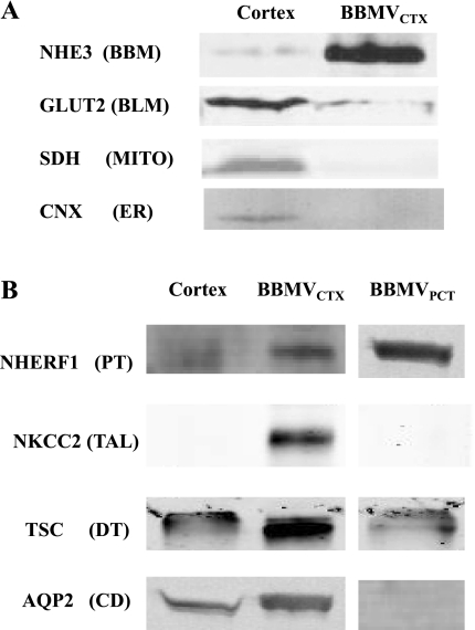
Fig. 2.
Western blot analyses of the purity of the BBMVCTX and BBMVPCT. A: analysis of contaminating organelle and basolateral membranes. Samples containing 20 μg of crude homogenates of rat renal cortex and BBMVCTX were separated by SDS-PAGE and probed with antibodies that are specific for the apical (BBM) Na+/H+ exchanger-3 (NHE3), the basolateral (BLM) glucose transporter (GLUT2), the mitochondrial (MITO) succinate dehydrogenase (SDH), and the endoplasmic reticulum (ER) marker calnexin (CNX). B: analysis of contaminating apical membranes. Samples containing 20 μg of renal cortex and BBMVCTX or isolated BBMVPCT were probed for proteins that, within the cortex, are expressed only in the proximal tubule (PT), Na+/H+ exchanger regulatory factor-1 (NHERF1); the thick ascending limb (TAL), Na+K+2Cl−-cotransporter-2 (NKCC2); the distal tubule (DT), thiazide-sensitive Na+Cl− co-transporter (TSC); and the collecting duct (CD), aquaporin-2 (AQP2). For each protein, the spacing denotes where a lane was deleted from the original figure of a single gel.

