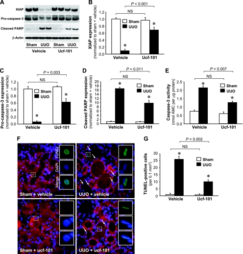
Fig. 6.
Effect of ucf-101, an inhibitor of Omi, on apoptotic signal activation and apoptosis induced by UUO. Male BALB/c mice were treated with either vehicle or ucf-101 for 5 days, beginning 2 days before UUO or sham. Kidneys were harvested 3 days after the operation. A: XIAP, pro-caspase-3, and cleaved PARP protein levels in the whole kidney lysate were determined via Western blot analysis using antibodies against these proteins, with β-actin used as an equal loading marker. B–D: protein bands were quantified using LabWorks analysis software. E: caspase-3 activity in the whole kidney lysate was measured using a Caspase-3 Fluorescent Assay Kit. Ac-DEVC-AMC was used a substrate of the caspase-3 enzyme. F: apoptosis was determined via TUNEL assay. Arrows indicate TUNEL-positive cells. White rectangular areas were separated into TUNEL, DAPI, and merged images. G: TUNEL-positive cells were quantified as the number of positive nuclei in a 0.1-mm2 field of the kidney section (10 fields/kidney). Blue indicates DAPI-stained nuclei. Red emitted at 650–710 nm is used here to view the general morphology of the tissue without any treatment. Values are means ± SE (n = 4). Scale bars = 50 μm. *P < 0.05 vs. respective sham.

