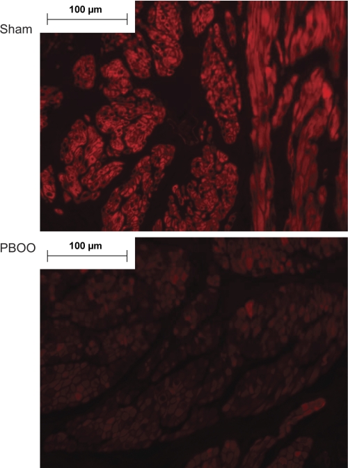
Fig. 3.
Confocal images of BKβ immunostaining in paraffin sections of bladders from rabbits after sham surgery or PBOO. Red fluorescent signal (Cy3) was used to detect BKβ, and the signal is much stronger in the sham sample than in the PBOO sample. Confocal images also show that BKβ is localized to smooth muscle cells. Nuclei are stained with by 4,6-diamidino-2-phenylindole (blue). Representative images are shown; n = 3.

