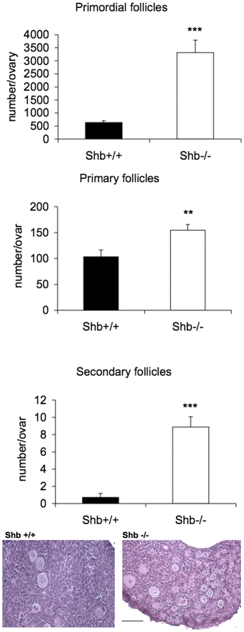
Figure 2. Follicle development of 18.5 dpc fetal ovaries from Shb +/+ and Shb −/− mice after culture for 7 days.
Fetal ovaries were cultured for 7 days and the organ explants morphology examined. Representative images are included in the figure (scale bar indicates 100 µm). Means ± SEM are given for n = 6–8. ** and *** indicate p<0.01 and <0.001, respectively compared with Shb +/+ using a Student's t-test or a Mann-Whitney rank sum test.

