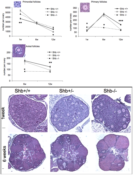
Figure 3. Number of primordial, primary and antral follicles from Shb+/+, Shb+/− and Shb−/− female mice at 1, 6 and 12 weeks of age.
Morphological analysis of ovaries at the different ages. Representative images are included (scale bars to the left indicate 50 µm). Values are means ± SEM (n = 10 except +/+ at week 1 where n = 7). * denotes a p<0.05; ** p<0.01; *** p<0.001 comparing Shb −/− with +/+ using ANOVA (Fisher LSD test at one and 12 weeks and Kruskal-Wallis variance on ranks at six weeks) or +/− primordial follicles at 12 weeks with +/+ (Kruskal-Wallis variance on ranks).

