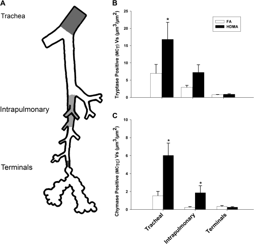
FIG. 3.
Abundance and distribution of MCT- and MCTC-positive cell (Vs) mass in specific regions in the airway wall. (A) The lung was microdissected open along the axial pathway, and airway branch points were marked. Trachea, intrapulmonary (generations 1–5), and terminal bronchioles (defined as proximal to the first alveolar outpocketings) were measured and compared. (B) MCT- and MCTC-positive cells were most abundant in the trachea and declined in the intrapulmonary and terminal airways. The trend is similar but exaggerated following HDMA exposure; both MCT- and MCTC-positive cell mass within the trachea was significantly greater than either intrapulmonary or terminal airway groups. Compared to FA controls, MCT cell mass was significantly increased in the trachea following the HDMA exposure regimen. (C) MCTC cell mass was significantly increased in both the trachea and the intrapulmonary airways. Terminal bronchiolar mast cells were not increased in response to HDMA exposure. Data are means ± SEM (n = 5–6 monkeys per group). *p < 0.05, as compared with FA controls in the same airway region.

