
Fig. 1.
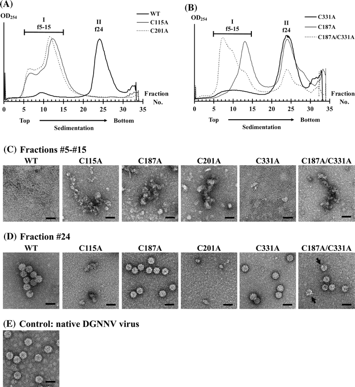
VLP formations from the DGNNV capsid protein with cysteine mutations. a The intensity profiles of wild-type and cysteine mutants determined by sucrose density gradient centrifugation. Bold line: WT (no mutation); thin line: C115A; dotted line: C201A. b Bold line: C331A; thin line: C187A; dotted line: C187A/C331A. Fractions from #5 to #15 are denoted as I-f5-15 and fraction #24 is denoted as II-f24. c, d Micrographs from fractions #5 to #15 and fraction #24 of the cysteine mutants. The arrow indicates the broken particles with irregular shape. e Control: native DGNNV virus. Bar: 50 nm