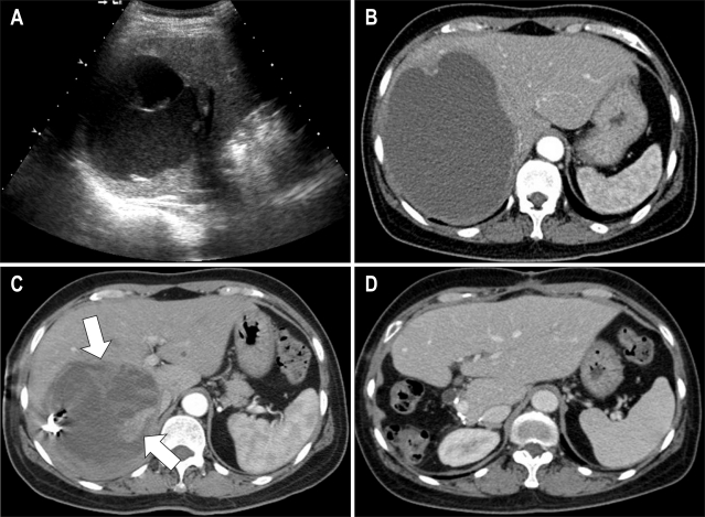
Fig. 1.
Undifferentiated liver sarcoma in a 53-year-old woman. (A) Ultrasonography showes an 8 cm cystic lesion with septation and echogenic material in the right lobe of the liver. (B) CT scan after 2 months reveales a hepatic cyst with a diameter that increases from 8 to 13 cm with slightly thickened periphery. (C) On follow-up CT performed 2 months after drainage, a solid lesion (arrow) mimicking granulation tissue is newly detected on the medial side of the cystic wall. (D) CT scan obtained 6 months after the operation showes no evidence of recurrence.

