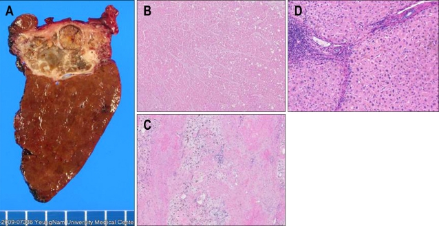
Fig. 3.
Pathology. A curative hepatectomy for segments 6 and 7 is performed. (A) Gross hepatectomy specimen showing necrotic liver mass. (B, C) Microscopic findings of the liver mass showing complete necrosis without any viable tumor cells (H&E stain, ×40). (D) Microscopic finding of the nontumor portion shows cirrhotic change (H&E stain, ×100).

